Executive Summary
If you're deciding between CTCs and ctDNA for your liquid biopsy research, here's what you need to know.
Both come from a simple blood draw. Both provide tumor-derived information without surgical biopsy. But they carry fundamentally different types of information, which means they're each better suited to different research questions.
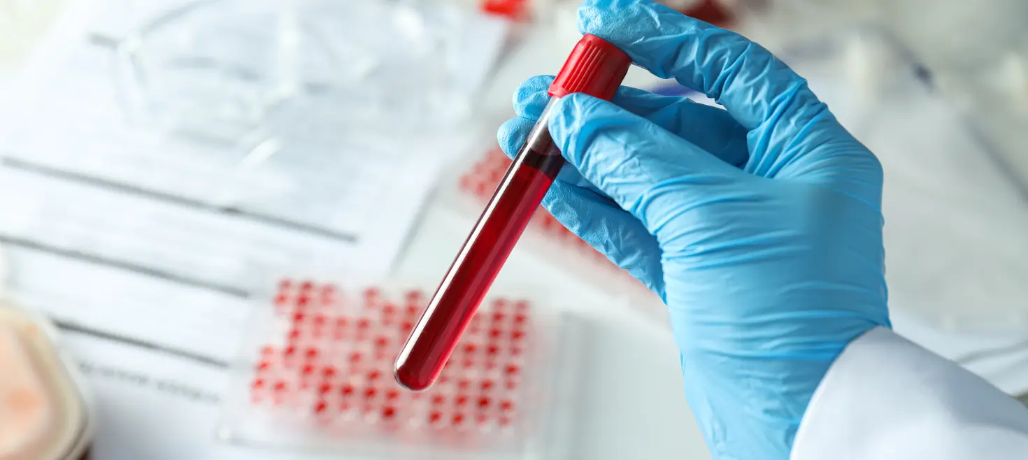
CTCs are intact, living tumor cells that escaped into the bloodstream. They can be cultured, tested for drug sensitivity, and analyzed for protein expression. You get whole cells with complete genomic material and functional proteins.
ctDNA consists of short DNA fragments released when tumor cells die. It enables sensitive mutation detection, methylation profiling, and rapid treatment response monitoring - but you lose all cellular context and protein-level information.
The key difference: CTCs tell you about viable tumor cells circulating right now. ctDNA tells you about tumor cells that recently died. Neither is better. The right choice depends on what you're trying to learn.
When cancer type matters: ctDNA detection rates exceed 75% for most metastatic solid tumors. But for glioblastoma (10-50% detection), renal cell carcinoma, and some prostate cancer contexts, ctDNA approaches struggle. CTCs become the more viable option.
What the combined data shows: In triple-negative breast cancer, using CTCs alone predicted recurrence with 62% sensitivity. ctDNA alone achieved 79%. Using both together reached 90%. The analytes capture different biology - combining them provides discriminatory power that neither achieves alone.
Key Takeaways
 CTCs provide living cells; ctDNA provides DNA fragments
If you need to culture cells, test drug sensitivity, or analyze protein expression, you need CTCs. If you need mutation profiling or methylation analysis, you need ctDNA.
ctDNA detection varies dramatically by cancer type.
High-shedding tumors (colorectal, pancreatic, lung) show >75% detection rates. Low-shedding tumors (glioblastoma at 10-50%, renal cell carcinoma, thyroid) may require CTC-based approaches.
EpCAM-based CTC capture misses the most dangerous cells
Over 73% of CTCs in breast cancer show mesenchymal features. Cells undergoing EMT - the ones driving metastasis - lose the markers that antibody-based methods need to catch them. Label-free capture methods solve this problem.
Combining CTCs and ctDNA improves sensitivity significantly
In triple-negative breast cancer, dual-analyte detection increased recurrence prediction from 62-79% (single analyte) to 90% (combined).
Pre-analytical requirements differ
ctDNA samples can be stable for 14 days in specialized tubes. Most CTC assays require same-day processing. Studies using both analytes typically need separate blood draws.
Neither analyte tells the complete story
CTCs may represent viable, treatment-resistant populations. ctDNA may preferentially capture dying cells responding to therapy. Together, they provide discriminatory capacity that neither achieves alone.
How do you get the best data for your work?
Liquid biopsy has become a practical reality for cancer research. But choosing between circulating tumor cells and circulating tumor DNA is not a question of which is better. It's a question about which one provides the best data for your work.
These two analytes come from different biological processes, carry different types of information, and perform differently depending on cancer type, disease stage, and what you're actually trying to learn.
What's the fundamental difference between CTCs and ctDNA?
The distinction starts at the source.
CTCs are intact, living tumor cells that have broken away from a tumor and entered the bloodstream through an active biological process - intravasation, often involving epithelial-mesenchymal transition. They contain complete genomic material, functional proteins, and intact RNA. They can be cultured, used for drug sensitivity testing, or analyzed at the single-cell level.
ctDNA comes from dead tumor cells. When tumor cells undergo apoptosis or necrosis, they release fragmented DNA into circulation. These fragments are characteristically short (around 134-144 base pairs) and turn over rapidly, with half-lives from 16 minutes to about 2.5 hours.
If you need living tumor material for functional studies, protein expression analysis, or drug sensitivity testing, you need CTCs. If you need sensitive mutation detection, methylation profiling, or rapid treatment response assessment, ctDNA is typically more practical.
How do detection methods differ?
CTC Isolation. Two Approaches
Marker-based capture uses antibodies against tumor-specific surface proteins:
- CellSearch (FDA-cleared) uses anti-EpCAM antibodies.
- Well-standardized with established prognostic thresholds.
- Cannot capture cells that have undergone EMT and lost epithelial markers.
Label-free capture uses physical properties instead of markers:
- Size-based filtration (ISET) uses membrane filters with 8 μm pores.
- Microfluidic systems (Parsortix) trap cells based on size and deformability.
- Captures both epithelial and mesenchymal CTCs.
- Less standardized but captures phenotypically diverse populations.
ctDNA Detection: Targeted vs. Broad
Which cancers shed enough ctDNA for reliable detection?
This is where cancer type becomes a critical variable. Detection rates vary dramatically:
High ctDNA Shedding (>75% detection in advanced disease)
- Small cell lung cancer (91%).
- Pancreatic cancer.
- Ovarian cancer.
- Colorectal cancer.
- Bladder cancer.
- Gastroesophageal cancer.
- Breast cancer.
- Melanoma.
- Hepatocellular carcinoma.
Low ctDNA Shedding (<50% detection)
- Glioblastoma: 10-50% - The blood-brain barrier blocks DNA release. This is biological, not technical - no assay improvement will fix it.
- Renal cell carcinoma.
- Prostate cancer.
- Thyroid cancer.
What This Means for Brain Tumor Research
For glioblastoma specifically, you have two options:

Cerebrospinal fluid sampling
achieves ~92% ctDNA detection, but more invasive

CTC analysis
CTCs detected in 77-84% of glioblastoma patients using size-based isolation.
For brain tumors, CTCs may be your only viable blood-based option.
Why do EpCAM-based methods miss the most dangerous CTCs?
This is arguably the most important technical consideration for CTC research, and it's underappreciated.
When tumor cells undergo epithelial-mesenchymal transition (EMT), they:

Downregulate
EpCAM, E-cadherin, cytokeratins

Upregulate
Vimentin, N-cadherin, Twist
EpCAM-based capture methods are blind to cells that have completed this transition. And the evidence suggests these are the cells that matter most.
The Numbers Are Striking
Bottom line: If your research focuses on metastatic mechanisms, EMT dynamics, or treatment-resistant populations, label-free capture methods aren't optional - they're essential.
What can CTCs tell you that ctDNA cannot?
CTCs provide access to information that is fundamentally inaccessible through cell-free DNA:
 Functional Studies
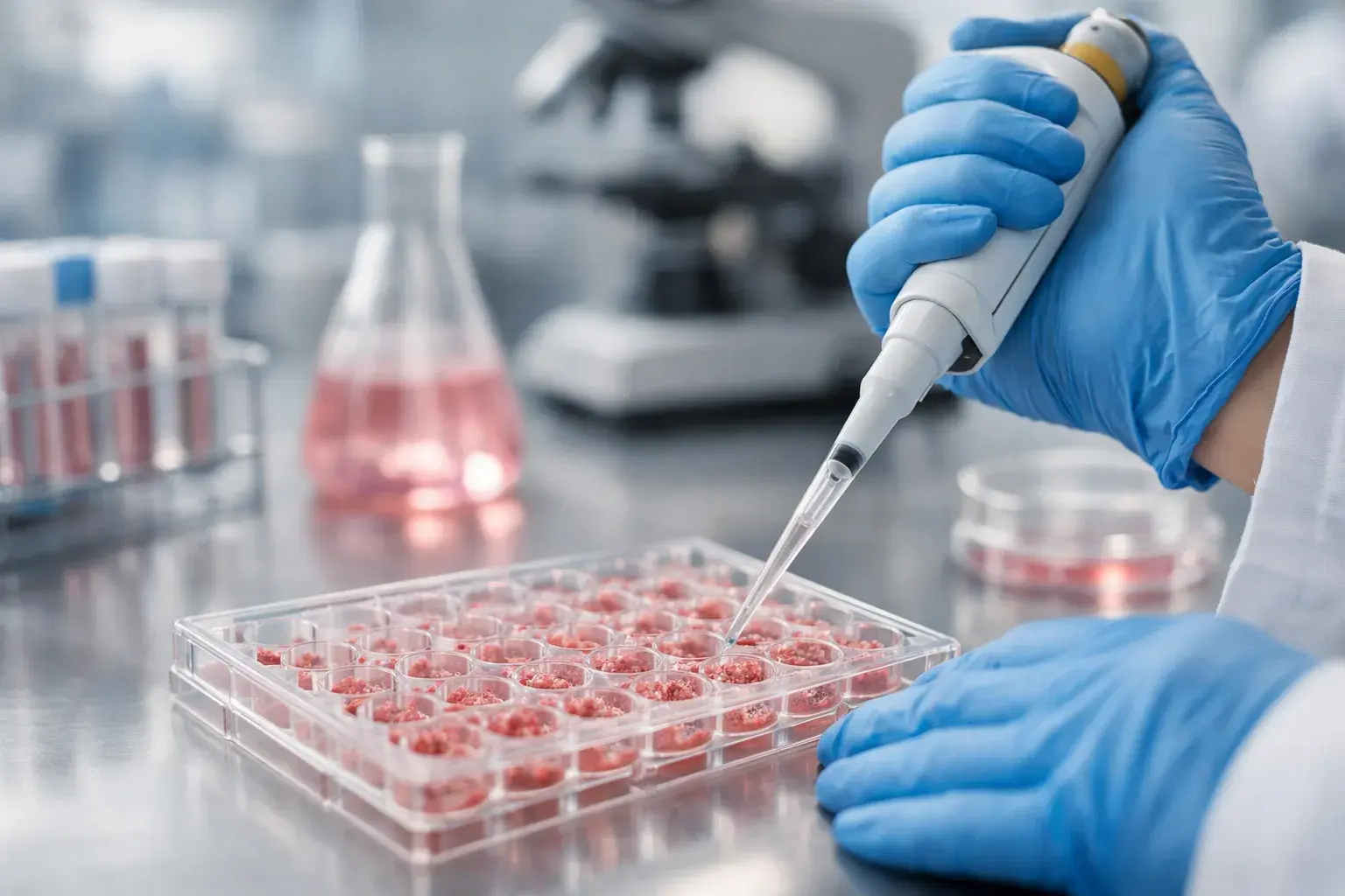
- Drug sensitivity testing - CTC culture has been achieved across breast, prostate, colon, lung, melanoma, and pancreatic cancers.
- Synergy discovery - One breast cancer CTC line with PIK3CA and FGFR2 mutations showed combined pathway vulnerability unpredictable from genomics alone.
- CTC-derived xenografts - Patient-specific models for therapeutic hypothesis testing (~5% success rate from standard draws).
 Protein Expression
- HER2 status can differ between primary tumor, metastases, and CTCs.
- PD-L1 expression on CTCs may predict immunotherapy response.
- Real-time pharmacodynamic readouts through surface marker monitoring during treatment.
 Unique CTC Phenomena
- Single-cell heterogeneity - RNA-seq of individual CTCs reveals immune evasion mechanisms (e.g., HLA-E:CD94-NKG2A axis).
- CTC clusters - Aggregates of 2-50 cells with dramatically enhanced metastatic potential compared to single CTCs.
What can ctDNA tell you that CTCs cannot?
ctDNA has its own exclusive capabilities.
 Methylation and Tissue-of-Origin
- CancerSEEK achieved 83% accuracy identifying tumor tissue of origin using methylation signatures.
- Tissue-specific patterns guide treatment when primary site is unknown.
 Fragmentomics
Analysis of fragment lengths, end motifs, and nucleosome positioning enables:
- Cancer detection without requiring mutations.
- Tumor subtyping.
- Gene expression inference.
 Minimal Residual Disease
- Post-surgical ctDNA positivity consistently predicts recurrence across colorectal, lung, and breast cancers.
- At low tumor burdens, ctDNA's higher relative abundance provides statistical advantages over rare CTC detection.
 Rapid Treatment Response
- The 16-minute to 2.5-hour half-life means you can see therapy response in days, not weeks.
- Declining ctDNA levels indicate response long before imaging changes.
Does combining CTCs and ctDNA improve results?
Yes - and the improvement is substantial, not marginal.
BRE12-158 Trial (Early-Stage Triple-Negative Breast Cancer)
Patients positive for both markers showed hazard ratios of 5.29 for distant disease-free survival.
COMET Study (Metastatic Breast Cancer)
- Only 28% overlap in mutations identified between CTC and ctDNA fractions.
- Optimal prognostic model combined baseline ctDNA VAF with CTC counts at four weeks.
- Neither analyte alone matched the combined model's performance.
EGFR T790M Detection (Lung Cancer)
Using both blood-based assays enabled successful genotyping in all patients - including T790M identification in 35% of cases where tissue biopsy was negative or indeterminate.
Why the complementarity? CTCs represent viable, potentially treatment-resistant cells. ctDNA may preferentially capture dying cells responding to therapy. Neither tells the complete story.
What practical factors should guide your decision?
Pre-Analytical Requirements
No single tube is validated for simultaneous CTC and ctDNA analysis. Dual-analyte studies typically require separate draws.
Throughput and Scalability
- ctDNA assays are generally more scalable for large studies.
- CTC isolation requires more hands-on time per sample.
- Automation is improving but not yet equivalent.
How should you match analyte to research question?
Choose CTCs When:
- You need living tumor material for functional studies or drug sensitivity testing.
- You're investigating metastatic mechanisms at the cellular level.
- Protein expression or surface marker profiling is central to your question.
- You're working with low-shedding tumors (glioblastoma, RCC, some prostate contexts).
- EMT biology or mesenchymal phenotypes matter (use label-free capture).
- Single-cell heterogeneity is important.
Choose ctDNA When:
- Comprehensive mutation profiling is the primary objective.
- You need maximum sensitivity for minimal residual disease.
- Rapid treatment response assessment matters (days, not weeks).
- Methylation analysis or tissue-of-origin is relevant.
- You're running large-scale studies prioritizing throughput and standardization.
- You're working with high-shedding tumors (>75% detection rates).
Consider Both When:
- Maximum prognostic or predictive accuracy is the goal.
- You're studying treatment resistance (CTCs = survivors, ctDNA = responders).
- Mechanisms manifest differently at cellular vs. molecular levels.
- The stakes justify the added complexity.
What does CTC analysis require in terms of workflow?
The practical workflow deserves attention if you're considering CTC-based approaches.
The Core Challenge
You're looking for a handful of cells among billions - and you need to:
- Capture them intact.
- Identify them reliably.
- Retain them through downstream analysis.
Where Cells Get Lost
The math: If you capture 10 CTCs but lose 70% during handling, you have 3 cells for analysis. Your data quality suffers accordingly.
The process in a nutshell

Label-free capture (size/deformability-based)
Catches epithelial AND mesenchymal CTCs.

High-retention slide system
77% retention vs. 30% for standard methods.

Multi-channel immunofluorescence
DAPI + epithelial markers + mesenchymal markers + CD45 to identify contaminating leukocytes.

EMT characterization
Distinguishes epithelial, mesenchymal, and hybrid phenotypes.
Where is the field heading?
The trajectory points toward multi-analyte integration, not declaring a winner.
Emerging Approaches
 Liquid biopsy troika
Combining CTCs, ctDNA, and extracellular vesicles for non-redundant information.
 Fragmentomics + AI
Machine learning on fragment patterns detects cancer without requiring specific mutations.
 Single-cell CTC technologies
RNA-seq and proteomics revealing immune evasion and resistance mechanisms.
 CTC-derived organoid
Patient-specific drug testing platforms maintaining individual tumor genetics.
 High-volume processing
Leukapheresis improving CTC yield for functional studies.
Researchers who develop competency with both CTCs and ctDNA will be best positioned as the field matures.
Yamato Life Sciences offers the CellBx Health liquid biopsy system, including the Parsortix PR1 for label-free CTC capture, CellKeep Slide for high-retention cell concentration, and Portrait+ immunofluorescence kit for CTC identification and EMT characterization. For researchers whose questions require intact tumor cells - particularly where EMT biology matters or ctDNA detection is limited - these tools address the practical challenges of CTC-based analysis.