Parsortix PR1
Harvesting rare CTCs as researchable “live cells”
It enables translational researchers to capture and harvest circulating tumor and other target cells from blood samples, offering enhanced flexibility for downstream analyses.
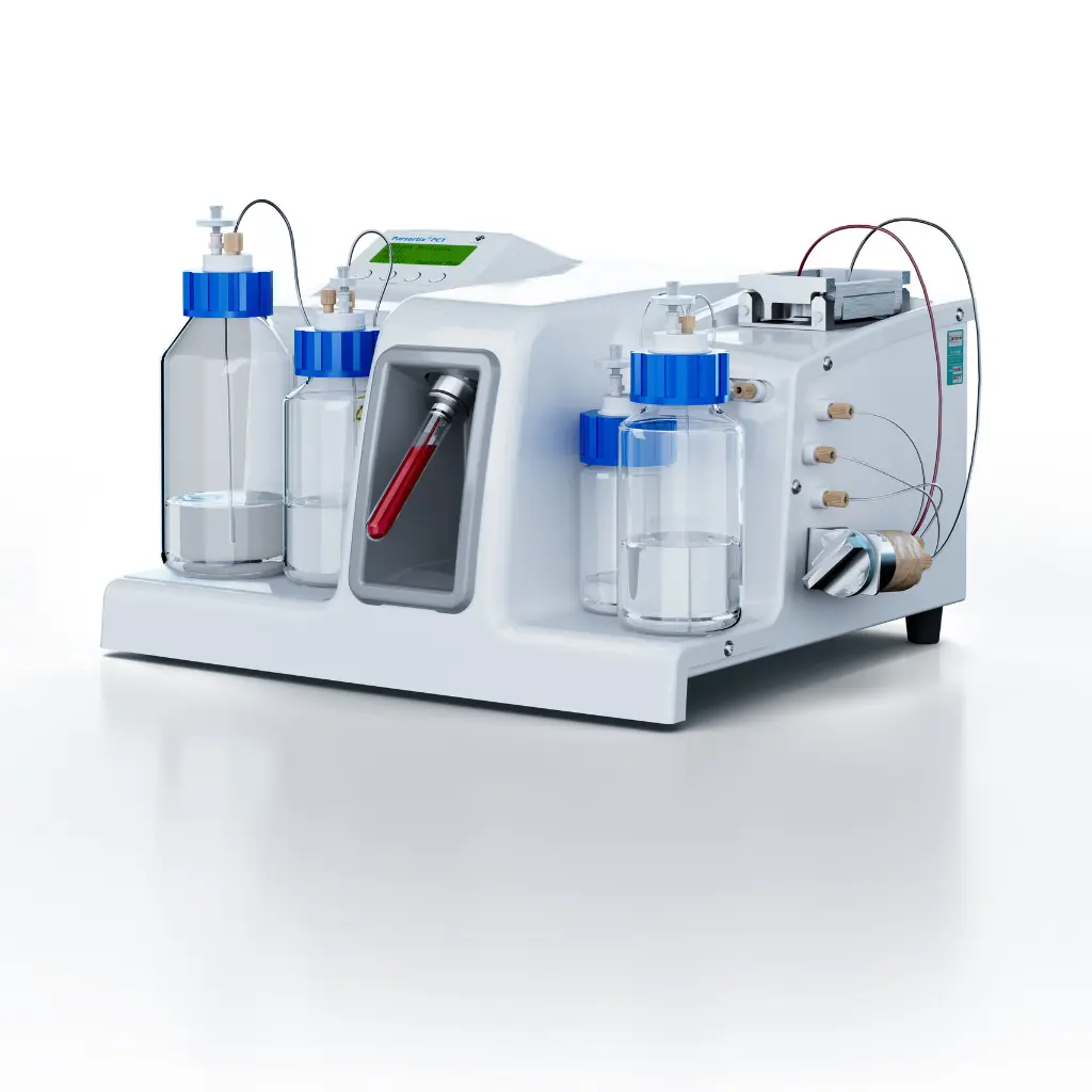
Parsortix PR1
A label-free CTC isolation system that recovers live, intact cells
Your capture method determines which cells you catch and what you can do with them afterward. Antibody-based platforms can’t see the EMT cells driving metastasis, and the cells they do catch come out dead. The PR1 catches the full CTC population and keeps every cell alive, so the research questions you set out to answer stay answerable.
The Parsortix PR1 is a semi-automated CTC isolation instrument that captures circulating tumor cells from whole blood based on their physical properties: size and deformability. Because capture depends on physics rather than antibodies or surface markers, the system isolates the full range of CTC phenotypes, including the EpCAM-negative and mesenchymal cells that antibody-based platforms cannot bind.
Captured cells come out alive. The system operates at under 0.5 psi, gentle enough to preserve viability for cell culture, drug sensitivity testing, single-cell sequencing, and other downstream work that requires living cells.
The PR1 handles circulating tumor cell isolation from sample volumes between 100 μL and 40 mL, drawn into standard EDTA blood collection tubes. No proprietary collection tubes required.
Label-free capture based on cell size and stiffness. No antibodies, no marker bias.
Cells come out alive, typically above 95% viability, ready for culture, drug testing, and sequencing.
Catches the EpCAM-negative and EMT cells antibody-based platforms miss: 41% recovery of EpCAM-low cells versus 0.35%.
Found CTCs in nearly twice as many lung cancer patients as the EpCAM-based standard (61% vs. 32%).
Runs on 100 μL to 40 mL of whole blood in standard EDTA tubes.
Depletes background blood cells up to 100,000-fold.
FDA De Novo cleared, with peer-reviewed publications across 19 cancer types.

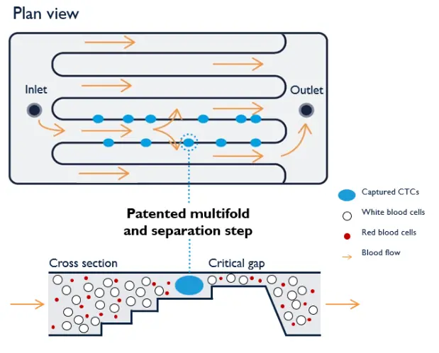
How CTC isolation works on the Parsortix PR1
Whole blood flows through a single-use microfluidic cassette containing serpentine channels that narrow in a series of steps. The final gap is 6.5 micrometers wide. Red blood cells (6-8 μm) and most white blood cells deform and pass through. Circulating tumor cells, which typically measure 12-25 μm and are stiffer than blood cells, are retained at the steps.
From there, you choose how to work with the captured cells...
Stain and image cells directly inside the cassette.
Reverse the flow and harvest cells into a 100-210 μL buffer volume for downstream analysis.
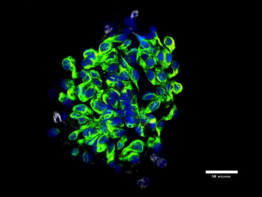
Because no capture antibody is involved, there is no phenotype bias. The same cassette retains epithelial CTCs, mesenchymal CTCs, hybrid phenotypes, and intact CTC clusters from a single blood sample, while depleting background blood cells up to 100,000-fold.
Parsortix technology is a unique method for capturing and harvesting intact circulating tumour cells (CTCs) and CTC clusters from whole blood for downstream analysis
CTCs are cancer cells that have detached from the primary tumour and entered the circulation. They are extremely rare in the blood and are often referred to as “a needle in a haystack”.
 As well as being functional cancer cells, CTCs play a critical role in initiating metastasis and are therefore a focus of cancer research and personalised medicine
By harvesting viable CTCs, Parsortix technology enables comprehensive profiling of cancer cells in a non-invasive, repeatable manner
Parsortix® technology uses a patented microfluidic technology in the form of a single use cassette to capture and then harvest CTCs from whole blood. The cassette captures CTCs based on their less deformable nature and larger size compared to other blood cells
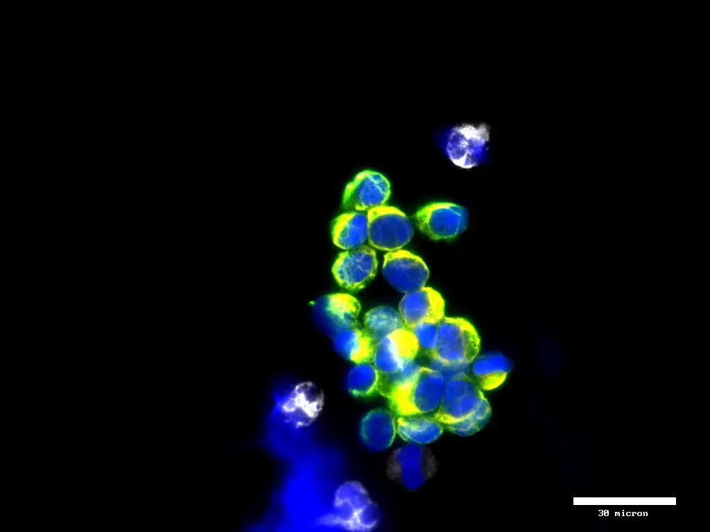
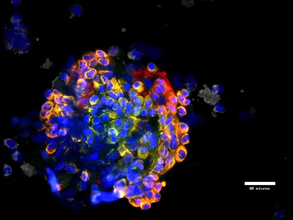
Why label-free CTC enrichment catches cells antibody-based methods miss
Antibody-based CTC capture depends on EpCAM, a surface protein expressed on epithelial cells. When cancer cells undergo epithelial-to-mesenchymal transition (EMT), they downregulate EpCAM. The cells most associated with metastasis and treatment resistance become invisible to the platforms built to find them.
In a breast cancer study using label-free isolation, 83% of CTC-positive patients had EpCAM-negative or mixed CTC populations that antibody-only methods would undercount or miss entirely.
In non-small cell lung cancer, size-based isolation detected CTCs in 61% of patients versus 32% for the EpCAM-based standard.
In matched prostate cancer samples, this Parsortix system captured 3.2 times more CTCs per sample than antibody-based capture.
When EpCAM-low cells were spiked into blood samples, the Parsortix system recovered 41% of them versus 0.35% for antibody-based capture.
Physical CTC enrichment sidesteps marker dependence entirely. Even after cells shed their epithelial markers, they remain larger and stiffer than blood cells, so the cassette still retains them.
Research Workflow Flexibility
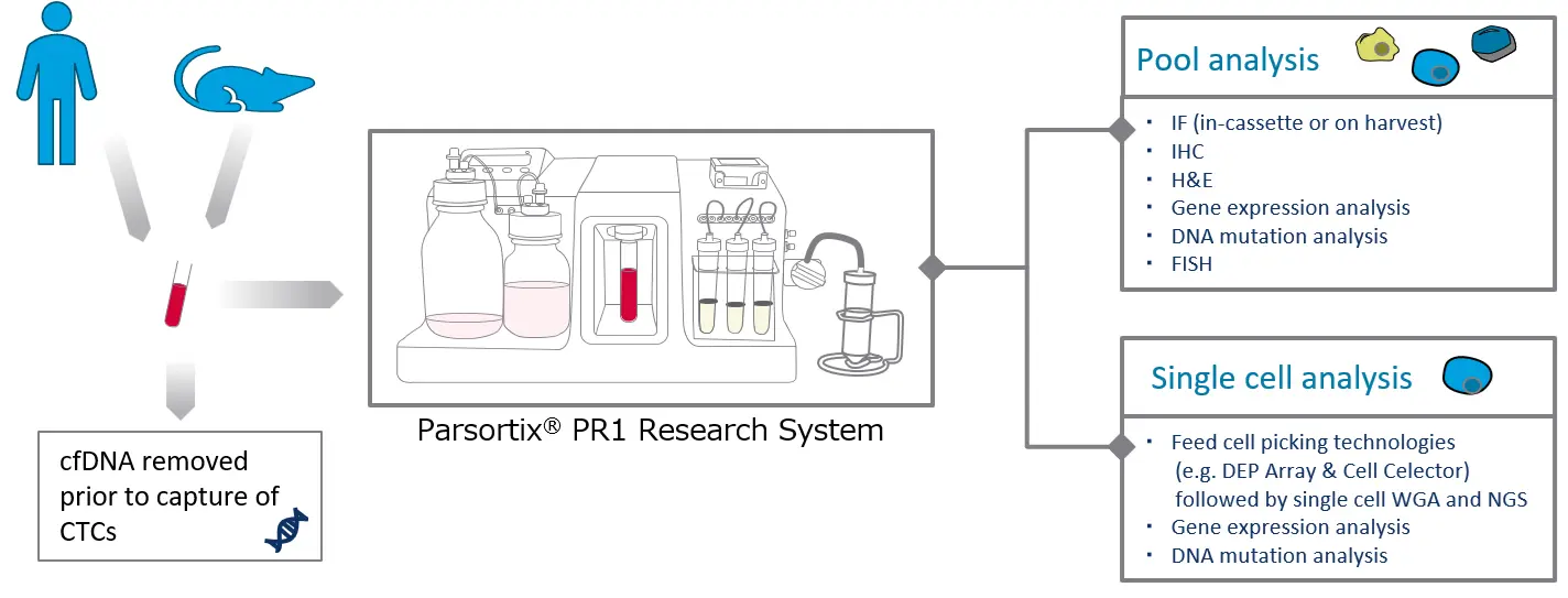
The Parsortix® PR1 Research System is designed to prioritize flexibility in downstream analysis following CTC recovery.
Pooled analysis
Immunofluorescence (IF), IHC, H&E staining, gene expression analysis, DNA mutation analysis), FISH
Single-cell analysis
Single-cell picking (e.g. DEPArray, CellCelector), single-cell WGA, NGS, gene expression analysis, and mutation analysis.
* It is also possible to implement a workflow that isolates ctDNA prior to CTC capture.
You may choose between *in vitro* staining or cell recovery to facilitate downstream analysis tailored to your specific research objectives.
CTC isolation instrument specifications
Health conditions investigated
The Parsortix PR1 system captures cancer cells from blood, as well as foetal cells from maternal blood. Peer reviewed publications evidence the efficacy of the system with a wide range of cancer types including:
Breast
Ovarian
Lung
Prostate
Melanoma
Pancreatic
Head and Neck
Gastric
Adrenal cortical carcinoma
Cervical
Colorectal
Brain
Renal
Endometrial
Sarcoma
Esophageal
Bladder
Neuroendocrine
Vulvar
What you can do with the cells after circulating tumor cell isolation
Fixed-cell platforms end at enumeration and imaging. Because the PR1 harvests viable, intact cells, the downstream menu opens up considerably...
CTC culture and expansion.
Drug sensitivity testing on a patient’s own circulating cells.
Patient-derived xenograft and CTC-derived explant models.
Single-cell RNA sequencing, whole genome amplification, and NGS.
DNA mutation analysis and gene expression profiling.
FISH, qPCR, IHC, and immunofluorescence.
Enumeration.
A workflow that isolates ctDNA from the same sample prior to CTC capture is also possible, so one blood draw can feed both analytes.
FDA clearance and published validation
This CTC isolation system received FDA De Novo clearance in 2022 for harvesting CTCs from metastatic breast cancer patients for subsequent analysis. The submission included data from more than 16,000 processed samples and was supported by the multi-center ANG-002 HOMING study.
Beyond the clearance, peer-reviewed publications document CTC isolation across 19 cancer types, including breast, lung, prostate, colorectal, ovarian, renal, brain, melanoma, sarcoma, and pancreatic cancers, along with fetal cell capture from maternal blood.
Which labs get the most from the Parsortix PR1
The PR1 fits research programs where marker-independent capture and live cells decide whether the work is possible at all.

Translational researchers studying metastasis, EMT, and treatment resistance, where the cells of interest lose the markers antibody platforms depend on.
Labs working with low-EpCAM cancers: triple-negative breast cancer, renal cell carcinoma, melanoma, and sarcoma.
Groups whose downstream work requires living cells for culture, drug testing, or xenograft models.
Teams running longitudinal monitoring studies that need flexible blood volumes across serial draws.
Researchers studying CTC clusters, which size-based capture retains intact.
Common questions about CTC isolation
CTC isolation is the process of separating circulating tumor cells from the normal cells in a blood sample. A blood draw from a metastatic cancer patient typically contains 1-10 CTCs per milliliter alongside billions of blood cells, so the isolation method has to enrich these rare cells enough to identify and analyze them.
The terms overlap and are often used interchangeably. Enrichment describes increasing the concentration of CTCs relative to background blood cells. Isolation describes recovering the cells themselves for analysis. The Parsortix PR1 does both: it enriches CTCs up to 100,000-fold over background blood cells and harvests them intact for downstream work.
Antibody-based systems capture cells by binding surface proteins, usually EpCAM. Cells that lose EpCAM expression during EMT go undetected. The Parsortix PR1 captures cells by size and deformability, so it retains epithelial, mesenchymal, and hybrid phenotypes plus CTC clusters, regardless of what markers the cells express.
Yes. The system runs at under 0.5 psi, and harvested cells typically show viability above 95%. That makes them suitable for culture, drug sensitivity testing, xenograft models, and sequencing applications where cell integrity affects data quality.
Anywhere from 100 μL to 40 mL, collected in standard EDTA tubes. Transfix tubes extend sample stability to 24-72 hours when same-day processing is not practical.
Peer-reviewed publications cover 19 cancer types, including breast, lung, colorectal, prostate, ovarian, renal, melanoma, brain, pancreatic, gastric, and head and neck cancers. The system also captures fetal cells from maternal blood.
Yes. The Parsortix system received FDA De Novo clearance in May 2022 for harvesting CTCs from metastatic breast cancer patients for subsequent analysis, backed by data from more than 16,000 processed samples.
Video
Publications

Nicolò, E. et al. (2025).
International expert consensus on the clinical integration of circulating tumor cells in solid tumors.
European Journal of Cancer, 231, 116050.

Bayou, N. et al. (2025).
Quantitative HER2 profiling on circulating tumor cells using an EpCAM-independent platform in metastatic breast cancer.
Cancer Cell Int. 25, 439.

Kurzeder, C. et al. (2025).
Digoxin for reduction of circulating tumor cell cluster size in metastatic breast cancer: a proof-of-concept trial.
Nat. Med. 1–5.

Obermayr, E. et al. (2024).
Gene expression markers in peripheral blood and outcome in patients with platinum‐resistant ovarian cancer: A study of the European GANNET53 consortium.
Int. J. Cancer ijc.34978

Davies, C. R. et al. (2023).
The potential of using circulating tumour cells and their gene expression to predict docetaxel response in metastatic prostate cancer.
Frontiers in oncology, 12.

Markou, A. N. et al. (2023).
Preoperative Mutational Analysis of Circulating Tumor Cells (CTCs) and Plasma-cfDNA Provides Complementary Information for Early Prediction of Relapse: A Pilot Study in Early-Stage.
Cancers, 15(6), 1877.

Ring, A. et al. (2022).
Circulating Tumor Cell Transcriptomics as Biopsy Surrogates in Metastatic Breast Cancer.
Ann. Surg. Oncol. 29, 2882–2894.