Skip to Content

Cellular assay using brightfield and fluorescence-based live cell imaging

Application Note: Cellular assay using brightfield and fluorescence-based live cell imaging using Celloger series to image cells in real time

This Application Note describes brightfield and fluorescence-based live cell imaging of HeLa cells using the Celloger platform to enable continuous, real-time imaging inside a standard incubator. Researchers face a persistent challenge in live cell analysis: capturing high-quality morphology and functional fluorescence data over long time courses without disrupting culture conditions or inducing phototoxicity. The Celloger solution addresses this problem through a compact, incubator-compatible design combined with contrast-enhanced brightfield optics and optimized low-intensity fluorescence pathways.

Developed and supported by Yamato Scientific America, Celloger enables brightfield live cell imaging for morphology monitoring and drug screening, as well as fluorescence-based live cell analysis for cytotoxicity and apoptosis assays, caspase-3/7 activation, and GFP transfection tracking. The system demonstrated superior brightfield contrast compared to comparable live cell imaging devices and fluorescence performance comparable to cMOS-based microscopes, while minimizing light-induced cellular stress. Real-time imaging of nocodazole-treated and staurosporine-treated cells revealed clear, quantifiable changes in confluency, cell death, and apoptotic signaling over time.

Together, these results position Celloger as a practical, high-performance solution for long-term live cell analysis in pharmacology and cell biology workflows.Download the full application note to explore the detailed protocol and comprehensive data set.


Live cell imaging technique makes it possible to understand and study various biological phenomena by enabling the observation of complex dynamics of live cells in real time using time-lapse microscopy. Realtime imaging of cellular phenomena such as cell migration, development and trafficking serves as an important means for research in various academic fields including cell biology, neuroscience, pharmacology and developmental biology. In order to observe the cells in a live state, incubator function is added to cover the microscope to control carbon dioxide, temperature and humidity (Figure 1A). But in many cases, controlling the temperature and humidity suitable for cell growth is challenging due to difficulties in maintaining airtightness and covering a large volume. To overcome such shortcomings, affordable and compact imaging devices that can be put into a cell culture incubator are being developed. Such live cell imaging devices basically provide bright-field images and at times come with fluorescence imaging functionality to observe fluorophores being excited and emitted in a specific wavelength. However, live cell imaging using fluorescence staining has a limitation since making fluorescence brighter and clearer not only results in improved image quality but inevitably causes cellular phototoxicity. Thus, it is essential for the time-lapse imaging system to enable efficient fluorescence imaging even at a low light intensity. As mentioned earlier, it is a crucial aspect for the live cell imaging system to ensure the image quality while maintaining temperature and humidity when processing experiments that generate significant amount of heat such as fluorescence imaging inside an incubator.

Celloger series, live cell imaging systems developed by Curiosis, are made in a compact size so that they can be placed in a general cell culture incubator (Figure 1B) and designed to endure the self-generated heat enabling the long-term imaging. In addition to that, it can obtain clear bright-field images using contrast-enhanced optics and fluorescence images of live cells in real time with a minimum light intensity by optimizing fluorescence filter and light path. The systems were tested to verify the applications in various cell-based research on the fields such as cell biology and pharmacology. The results showed that the devices had higher bright-field image quality than other live cell imaging system with the same functions and fluorescence imaging results were comparable to the images obtained from fluorescence microscopy using cMOS cameras with specifications corresponding to that of Celloger.


1. Bright-field imaging application

Drug screening is a very important and essential process for the development of drugs including anticancer drugs. For drug screening, it is important to obtain a clear image in the process of real-time cell monitoring while performing treatment according to the type or concentration of a drug. Celloger’s bright-field imaging has increased the contrast in comparison to the existing live cell imaging equipment, making it possible to display more vivid cell contours and boundaries despite the usage of transparent cell samples (Figure 2).


1.1. Morphology monitoring and drug screening

With Celloger Mini, the designated positions of multiple points can be scanned according to a set schedule as it has automatic motorized stages. This feature makes it possible to track the changes over time when cells are treated with different drug concentrations. Nocodazole, one of the anticancer drugs, is known to cause mitotic arrest by inhibiting the polymerization which cell and what concentration it is used1. Cells were treated with different concentration levels of nocodazole and observed by Celloger Mini. The results showed that most cells died had similar confluency at the final endpoint, 20 hours after the treatment with the drug when the drug concentration is over 62.5nM. On the contrary, there was difference in cell death and confluency depending on concentration levels of the drug in early time.

As such, it was possible to obtain important data for morphological dynamics of cells and antitumor efficacy of drug through real-time cell monitoring and confluency imaging using Celloger Mini. As shown in Figure 3, time-lapse images are generated in tile images, making it easy to compare the differences depending on conditions.


2. Fluorescence imaging application

Using live cell imaging equipment such as Celloger Nano, it becomes possible to visually investigate the dynamics of intracellular changes using the live cell staining fluorescent dyes with specific staining properties for subcellular organelles and cell labelling. Using this characteristic, it is possible to monitor and quantify the efficacy of a drug through various mechanisms. Fluorescence optics of Celloger Nano were optimized to increase the ratio of detected fluorescence to light source intensity, resulting in improved fluorescence image quality while minimizing phototoxicitythat occurs inevitably during excitation. The fluorescence images taken by Celloger Nano were compared with those taken by a fluorescence microscope equipped with a ASI174MM camera (SONY IMX174 cMOS image sensor) whose specification is comparable to that of Celloger Nano to verify the quality of fluorescence images. The fluorescence image of Hela cells stained with fluorescence dye using CMFDA, a green fluorescent cell tracker, taken by Celloger Nano showed that the fluorescence intensity was comparable to that of fluorescence microscope and the image was clear since the contrast between the background and cells was high (Figure 4).


2.1. Cytotoxicity assay

Several staining reagents that measure the degree of cell death using a phenomenon in which the integrity of the cell membrane is damaged and the cell permeability is increased during the cell death are commercially available. To measure the cytotoxicity by nocodazole, dead cells were stained with green fluorescent CellTox™ dye. It was confirmed that the number of cells measured by fluorescence increased as the cell permeability increases due to cell death after 20 hours (Figure 5).


2.2 Apoptosis assay

Fluorescence coverage graph is shown to quantify the apoptosis by time. The graph illustrates that fluorescence began to be detected from two and a half hours after the treatment with staurosporine and reaction became saturated from 10 hours after the treatment, making it possible to detect fluorescence in all cells (Figure 8).


2.3 Transfection


Conclusion

The Celloger series improves the efficiency of fluorescence imaging by enabling imaging even at a minimum level of excitation light, which can also reduce phototoxicity caused by fluorescence staining, a priority consideration for live cell imaging. The Celloger systems that were used to carry out the applications mentioned above work perfectly inside an incubator, which makes them ideal tools for various imaging applications and experiments.

Live cell imaging allows researchers to observe cellular processes such as migration, division, and death in real time. This approach provides dynamic information that endpoint assays cannot capture, making it valuable in cell biology, pharmacology, and drug discovery.

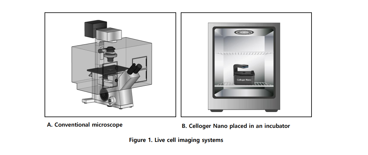
Bright-field imaging visualizes cells based on differences in light transmission through the sample. The Celloger system uses contrast-enhanced optics to clearly define cell contours and boundaries, even for transparent live cells, without the need for staining.

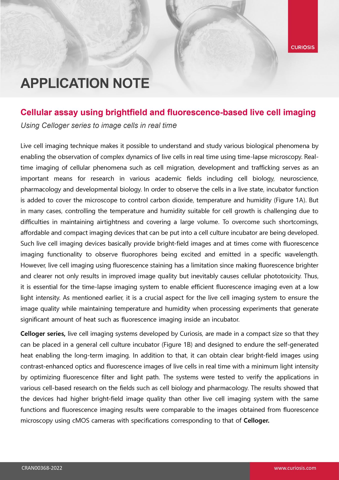
Celloger instruments are compact and designed to operate directly inside standard incubators while maintaining temperature, humidity, and CO₂ conditions. They are engineered to withstand self-generated heat and still deliver stable, high-quality images during long-term time-lapse experiments.

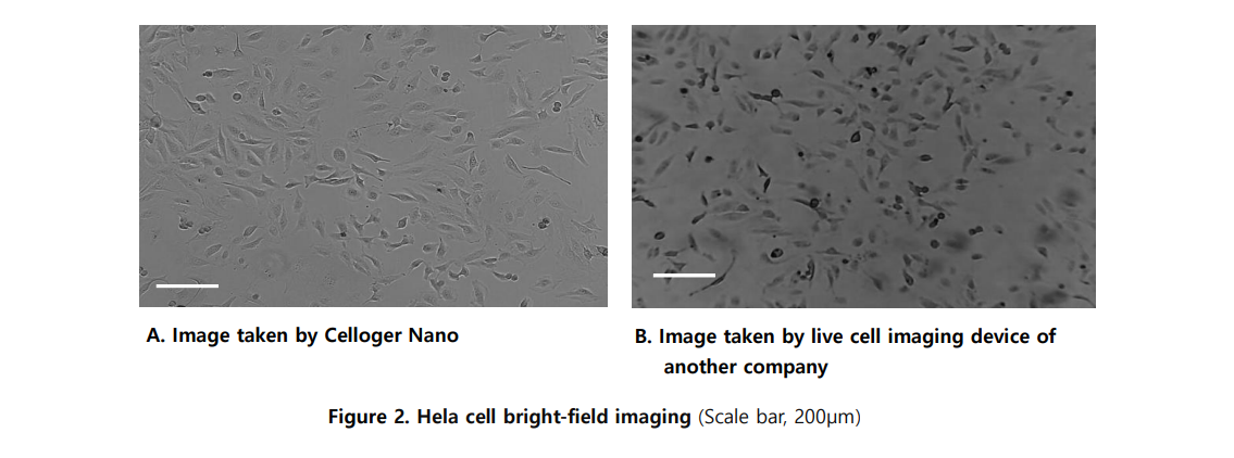
In the application note, bright-field drug screening experiments were conducted for 20 hours with hourly image acquisition. Fluorescence-based assays ran up to 40 hours, depending on the biological process being monitored.  

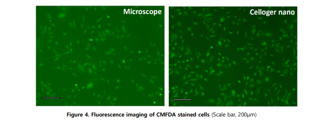
Celloger optimizes the fluorescence filter and light path to maximize detected signal at minimal excitation intensity. This design enables clear fluorescence imaging while reducing phototoxicity, which is critical for maintaining cell viability during long-term live imaging.

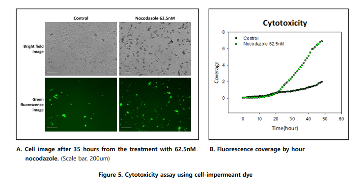
Yes, Celloger supports cytotoxicity assays using cell-impermeant fluorescent dyes such as CellTox™. The increase in fluorescence over time reflects loss of membrane integrity and allows real-time quantification of drug-induced cell death.  

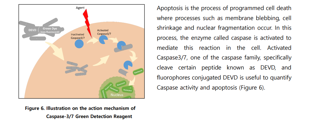
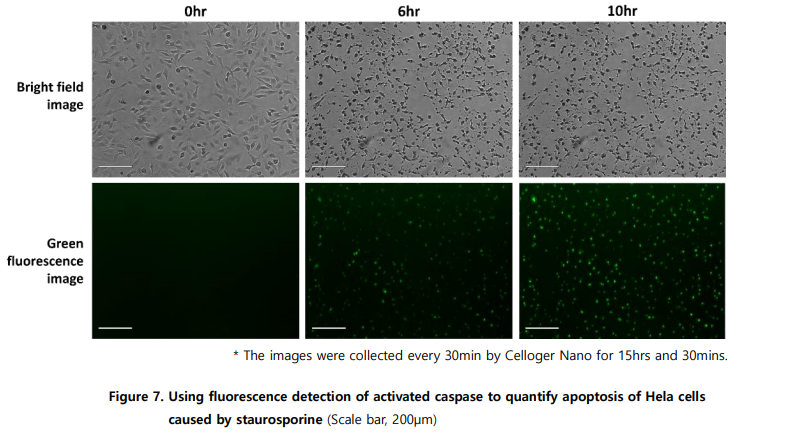
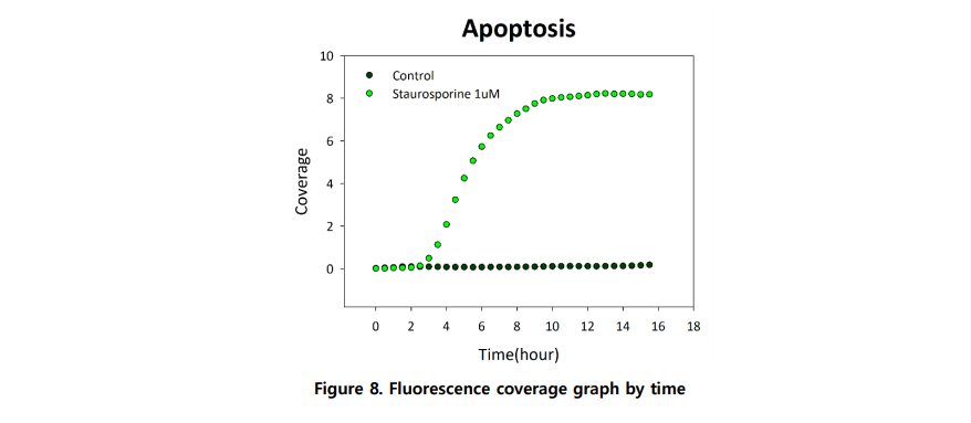
Apoptosis can be quantified using fluorophore-conjugated DEVD substrates that detect activated Caspase-3/7. Using Celloger Nano, fluorescence signaling apoptosis was detected within 2.5 hours of treatment and reached saturation around 10 hours.

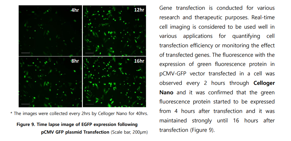
Yes, Celloger Nano enables real-time monitoring of fluorescent protein expression after transfection. In the application note, GFP expression was detected 4 hours post-transfection and remained strong for over 16 hours.  

The application note demonstrates successful imaging of HeLa cells across bright-field and fluorescence assays. Based on the described functionality, Celloger systems are suitable for a wide range of adherent live cell cultures used in cell biology and pharmacology research.

Download the full Application Note PDF

to access the complete brightfield and fluorescence live-cell imaging workflow, including incubator-based Celloger setup, HeLa cell protocols, contrast and fluorescence performance comparisons,

 and time-resolved analysis of drug-induced cytotoxicity and apoptosis.


Zebrafish observation using the Z stacking function