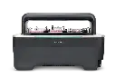
High-throughput live imaging system
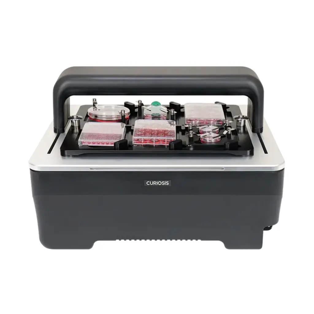
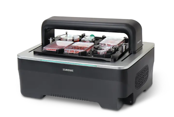
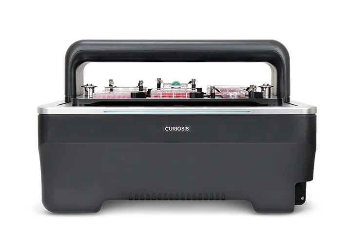
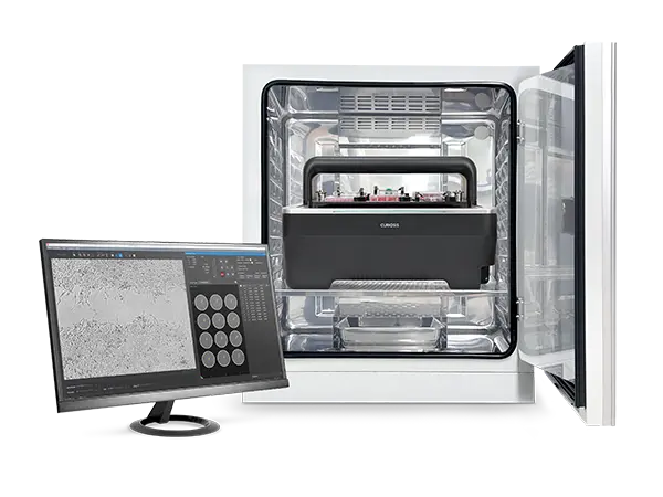
The Celloger® M26 is a next-generation high-throughput live cell imaging system designed to transform real-time cellular research. Engineered for performance, flexibility, and precision, the M26 delivers ultra high-resolution images with synchronized illumination to ensure uniform and reliable data across every well.Equipped with three user-interchangeable lenses and dual imaging modes—brightfield and fluorescence—the M26 enables comprehensive cellular analysis under various experimental conditions. Its compatibility with multiple plate formats and simultaneous imaging of up to six plates makes it an ideal solution for large-scale, uninterrupted cell monitoring inside your incubator.Whether you're conducting time-lapse studies, drug screening, or stem cell research, Celloger® M26 streamlines your workflow and elevates your imaging capabilities—without disrupting the cell environment.
Key Features

Ultra High-Resolution Imaging
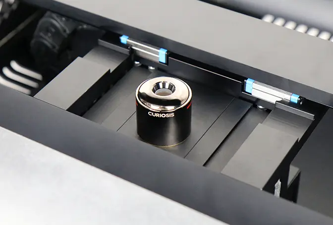
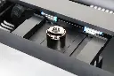
Interchangeable Lenses
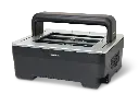
Multi-Plate Compatibility
Synchronized Illumination System
Brightfield & Fluorescence Modes
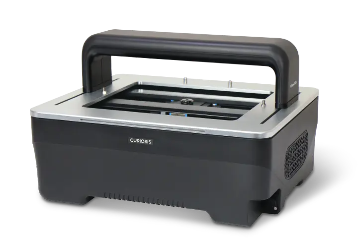
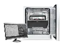
Incubator-Compatible Design
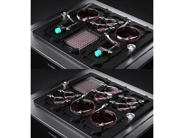
High-Throughput Multi-Plate Imaging
Capture up to six plates simultaneously under identical conditions, enabling reliable, reproducible, and scalable live-cell studies.
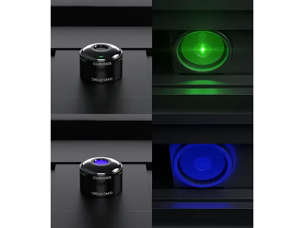
Synchronized Illumination System
Camera and light source move together to deliver consistent brightness and resolution across every position—ideal for precise time-lapse imaging.

Real-Time Monitoring in Incubators
Observe cell dynamics directly inside the incubator with single-cable connectivity, ensuring stable long-term experiments without disturbing culture conditions.