Skip to Content

Celloger® M22

A dual-vessel automated live cell imaging system designed for simultaneous real-time monitoring and comparison of cell cultures inside your incubator.

1.00 1.00

Not Available For Sale

    Please contact our team for help with this particular configuration here. Our expert team will respond to you very promptly.

    https://www.yamato-usa.com/web/image/product.template/14380/image_1920?unique=d50f019

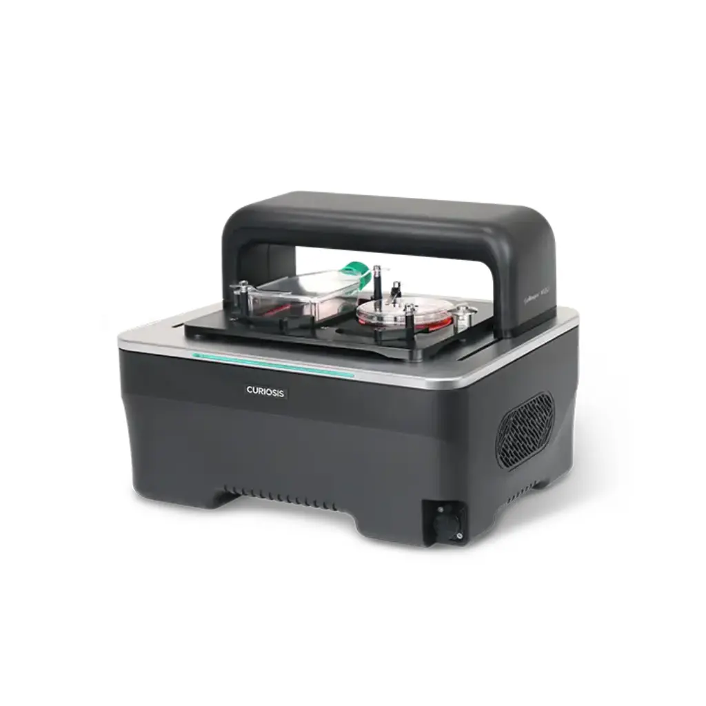
    Dual-vessel live cell imaging system

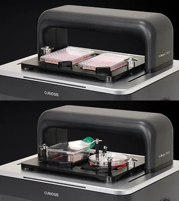
    Celloger® M22 is a dual-vessel live cell imaging system that enables simultaneous monitoring of two culture vessels inside the incubator.

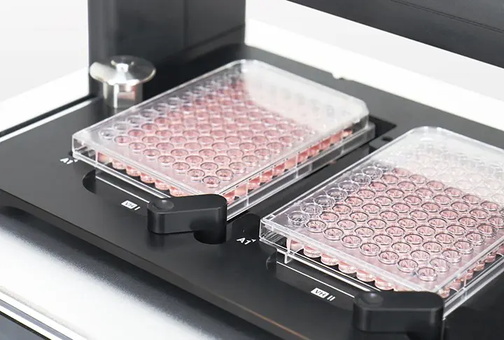
    By capturing both vessels in parallel with synchronized illumination, Celloger® M22 ensures consistent imaging conditions and minimizes variability during long-term observation.

    This configuration supports a wide range of experimental workflows, including control versus treatment studies, dose comparisons, and condition optimization, within a single, integrated imaging system.

    Key Features

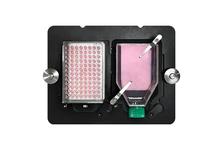
    Dual-vessel parallel imaging

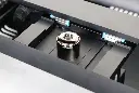
    User-interchangeable objective lens (4× / 10× / 20×)

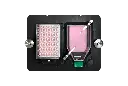
    Dual fluorescence & brightfield imaging

    Synchronized illumination for consistent imaging

    Temperature-stable operation

    Incubator-compatible design


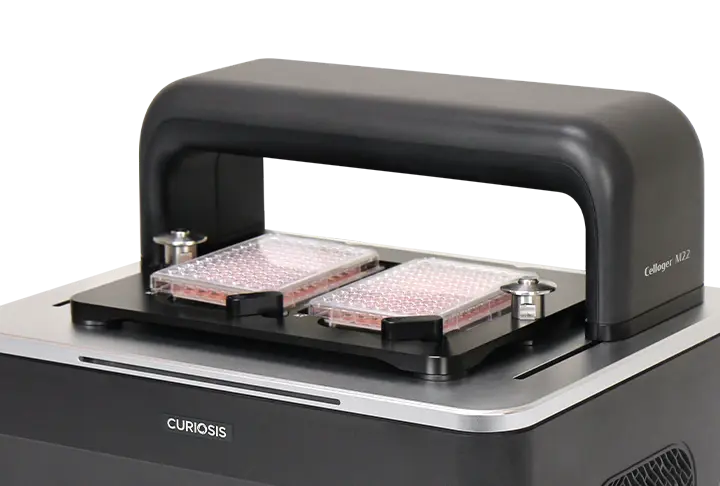
    Dual-Vessel Parallel Imaging

    Camera and light source move together to deliver consistent brightness and resolution across every position—ideal for precise time-lapse imaging.

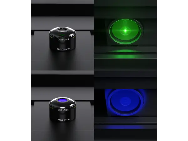
    Synchronized Illumination System

    Observe two culture vessels simultaneously within a single, synchronized imaging system. Celloger® M22 enables parallel imaging of two vessels under stable incubator conditions, supporting both identical and independent experimental setups.

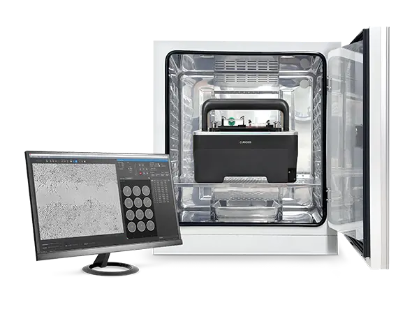
    Real-Time Monitoring in Incubators

    Observe cell dynamics directly inside the incubator with single-cable connectivity, ensuring stable long-term experiments without disturbing culture conditions.


    Specifications

    Imaging modes Brightfield, 2 fluorescence channels (Green & Red)
    Objectives 4× / 10× / 20× (user-interchangeable lenses)
    Fluorescence modes (customizable) Green (EX: 470/40, EM: 540/50)
    Red (EX: 562/40, EM: 641/75)
    Stage movement Fully motorized XYZ (camera moving type)
    Image sensor High sensitivity 5.0 MP CMOS
    Analysis methods Single/multicolor, stitching, Z-stacking, time-lapse, real-time recording
    Focusing Auto focusing
    Culture vessels Well plate up to 96-well, flask, dish, slide
    Software Scan App (acquisition), Analysis App (analysis)
    File export format TIFF, AVI (JPEG, PNG)
    Operating environment 10~40°C, 20~95% humidity
    Power supply 100-240V, ~50/60Hz
    PC requirements (O/S required) Windows 10 and above
    Incubator specification Above 170 L (recommended)
    Dimensions (mm) W:380 D:303 H:313
    Weight 10.86kg (main unit only, vessel holder excluded; 2-well plate holder: 0.85kg)