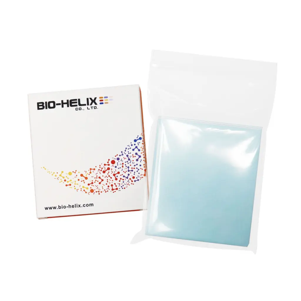
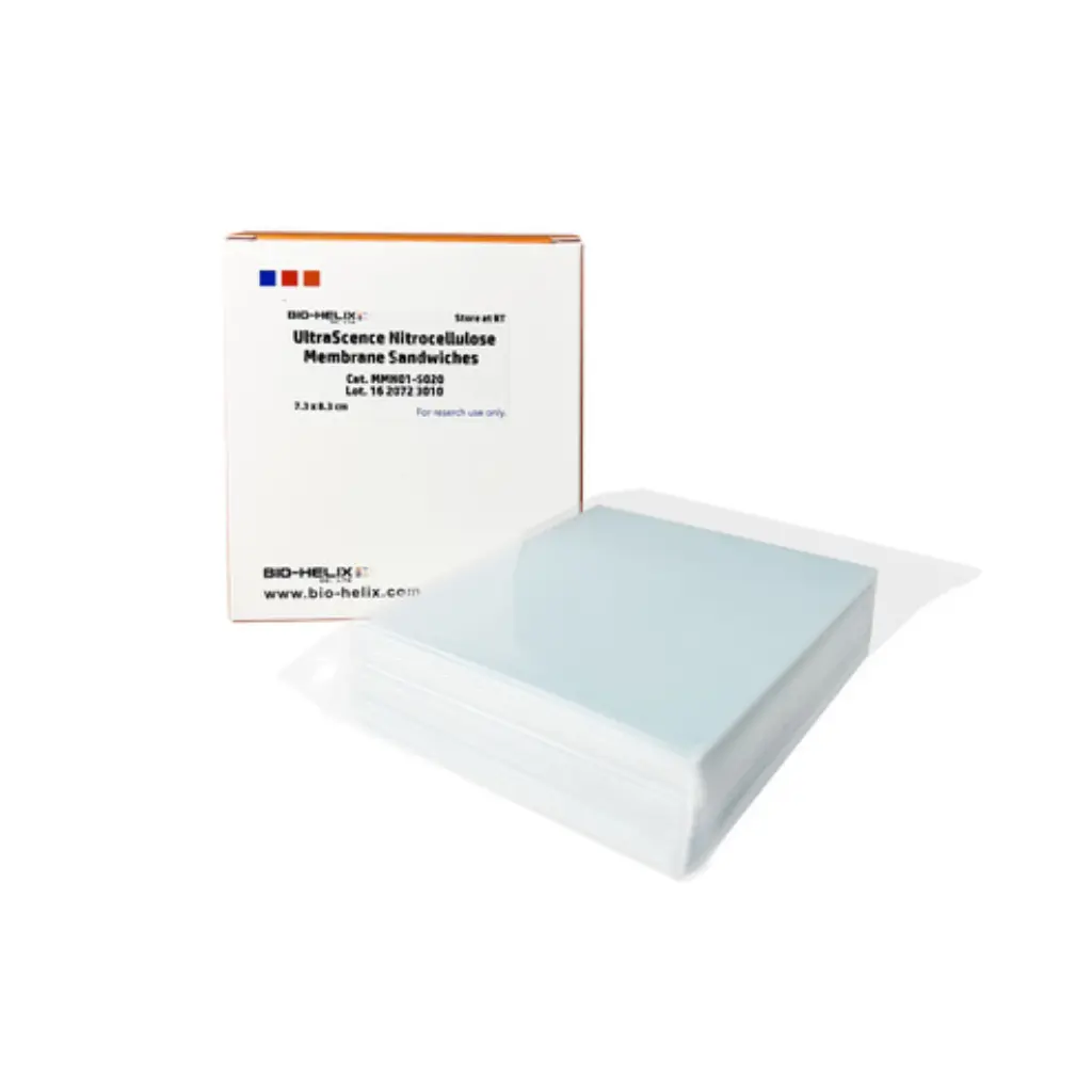
UltraScence Precut NC Membrane Sandwiches
BHMMN01S020
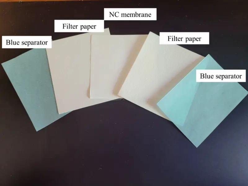
Nitrocellulose/Filter Paper Sandwiches, 0.2 μm, as high-quality membranes ideal for blotting proteins and nucleic acids, are ideal for the transfer of low molecular weight proteins (less than 20 kDa) and nucleic acids (less than 300 bp), exhibiting high sensitivity and low background for immunoblotting.
Specifications
*Incubation method with Glucose-6-phosphate dehydrogenase
Performance
Table 1. The protein binding capacity of the NC membrane.
The NC membranes were incubated in 0.5 mg/ml protein solution at room temperature for 24 hrs. Absorbance values were detected at 280 nm by the BCA assay, and the protein binding capacity can be calculated.
* PALL is a registered trademark of PALL Corporation. The trademark holder is not affiliated with Bio-HeliX Co., Ltd. and does not recognize this product.
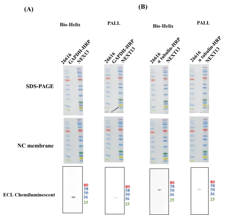
Figure 2. Western blotting image of NC membrane with UltraScence Femto Plus Western Substrate
Bio-Helix: Cat. MMN01-S020 / MMN02-S020
PALL: Cat. 66485
5 ug MCF-10A cell lysate was running in 4-20% Tris-Glycine gel. The protein bands were transfer to NC membrane by wet transfer (70V, 60 mins). The membrane was cut into two separate membrane for blocking. Blocked with One-step blocking buffer for 2 hrs. (A) membranes were probed with HRP-conjugated GAPDH Monoclonal antibody (Proteintech, 1:50,000) and HRP-conjugated affinipure Goat Anti-Mouse IgG secondary antibody (Proteintech, 1:100,000). (B) membranes were probed with Rabbit anti α-tubulin antibody (Abcam, 1:3000) and Goat Anti-Rabbit IgG/HRP secondary antibody (Solarbio, 1:10,000). The HRP-conjugated secondary antibody was applied and developed with UltraScence Femto Plus Western Substrate, and exposed for 2 seconds using Chemlux SPX-600 Series digital imaging system.
* PALL is a registered trademark of PALL Corporation. The trademark holder is not affiliated with Bio-Helix Co., Ltd. and does not recognize this product.
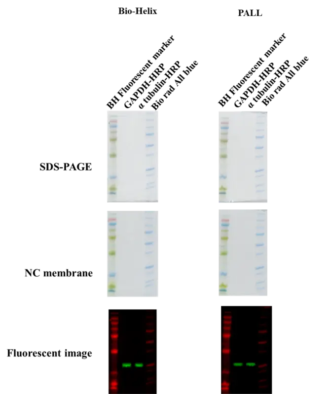
Figure 3. Fluorescent image of NC membrane
Bio-Helix: Cat. MMN01-S020 / MMN02-S020
PALL: Cat. 66485
5 ug MCF-10A cell lysate was running in 4-20% Tris-Glycine gel. The protein bands were transfer to NCmembrane by wet transfer (70V, 60 mins). The membrane was incubated with One-step blocking buffer for 2 hrs. (A) membranes were incubated with HRP-conjugated GAPDH Monoclonal antibody (Proteintech, 1:50,000) and Multi-rAb™ CoraLite® Plus 594-Goat Anti-Mouse Recombinant Secondary Antibody (H+L) (Proteintech, 1:50,000). (B) membranes were incubatedd with Rabbit anti α-tubulin antibody (Abcam, 1:3000) and Multi-rAb™ CoraLite® Plus 594-Goat Anti-Mouse Recombinant Secondary Antibody (H+L) (Proteintech, 1:50,000). Excitation: Green LED, emission: 565~625 nm, exposure time: 1 sec. Excitation: RED LED, emission: 700~740 nm, exposure time: 2 secs.
* PALL is a registered trademark of PALL Corporation. The trademark holder is not affiliated with Bio-Helix Co., Ltd. and does not recognize this product.
