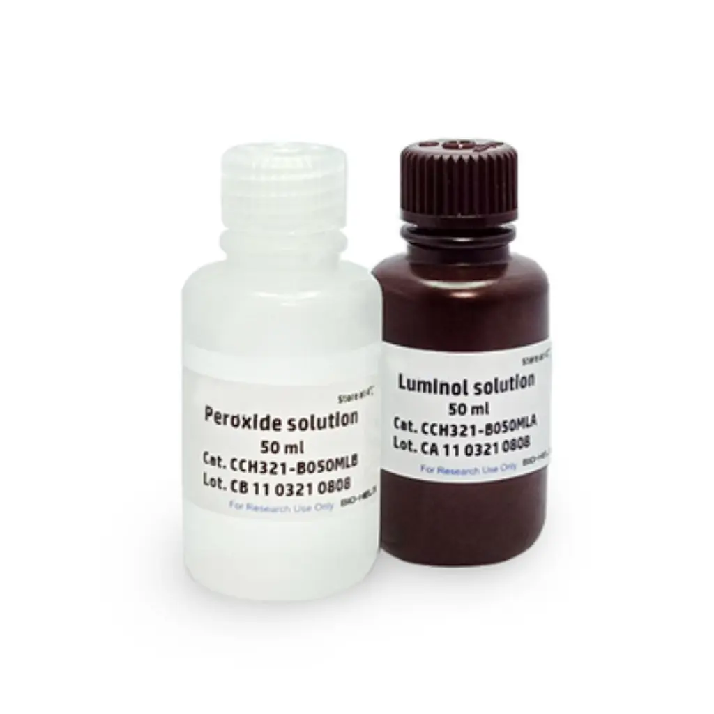
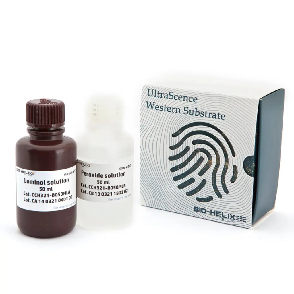
UltraScence Pico Plus Western Substrate
BHCCH321B100ML
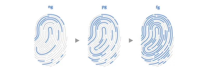
The UltraScence Pico Plus Western Substrate, as a luminol-based enhanced chemiluminescent substrate, is sensitive and compatible with conducting immunoblots with horseradish peroxidase (HRP) – conjugated secondary antibodies. The low picogram to high femtogram detection of antigen is enabled by UltraScence Pico Plus Western Substrate’s excellent sensitivity and long signal duration. Further, its long chemiluminescent signal duration makes both digital and film-based imaging possible without any loss of the signal. Appropriate primary and secondary antibody dilutions are suggested for attaining optimal signal intensity and duration.
Features

No optimization required
Switching to the UltraScence Pico Plus Western Substrate from other brands, such as Thermo Scientific™ SuperSignal™ Pico Plus and Bio-Rad™ Clarity™, does not require optimization or protocol changes.
High degree of sensitivity and enhanced chemiluminescence duration
UltraScence Pico Plus Western Substrate enables an accurate low picogram to high femtogram detection of protein on the same immunoblot after a single exposure.
Optimized for use with PVDF and nitrocellulose membranes
Compatible with Western Blotting Markers
Optimized for film and CCD-based imaging
Reaction Setup
1
Keep the membrane moist in the wash buffer while preparing the substrate mixture. Make sure the membrane does not dry out in the next steps.
2
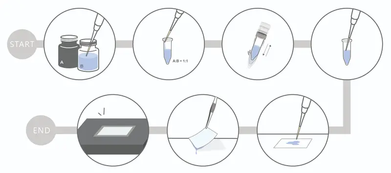
The chemiluminescent substrate solution is sufficiently stirred to prepare the 0.1ml of solution / cm2 of the membrane.
- For a small membrane (7 x 8.5 cm), 4 ml of solution is sufficient.
- For a medium-sized membrane (8.5 x 13.5 cm), 10 ml of solution is sufficient.
3
Place the membrane on a clean, flat surface or in a clean container.
4
Remove the membrane from the chemiluminescent substrate solution and drain the excess solution.
5
Place the membrane in a plastic sheet protector or plastic wrap to prevent the film from drying out.
6
Imaging the membrane with a digital imager or by exposure to the X-ray film.
Performance
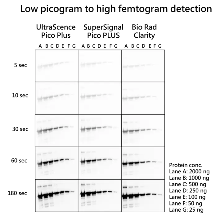
Figure 1. UltraScence Pico Plus Western Substrate enables an accurate low picogram to high femtogram detection of protein on the same immunoblot after a single exposure. Membranes were probed with GFP tag Rabbit PolyAb (Cat No. 50430-2-AP, 1:10,000) of and then with Goat Anti-rabbit IgG/HRP secondary antibody (Cat No. SA00001-2, 1:10,000) after serial dilution EGFP (Enhanced Green Fluorescent Protein) were prepared and applied in electrophoresis and protein transfer. Identical blots were incubated with the Western substrate. The blots were simultaneously exposed for 5 seconds, 10 seconds, 30 seconds, 60 seconds, and 180 seconds using ChemluxSPX-600 Series digital imaging system.
SuperSignal West Pico PlUS and Clarity Western ECL Substrate is a registered trademark of Thermo Fisher Scientific and Bio rad. The trademark holder is not affiliated with Bio-HeliX Co., Ltd. and does not recognize this product.
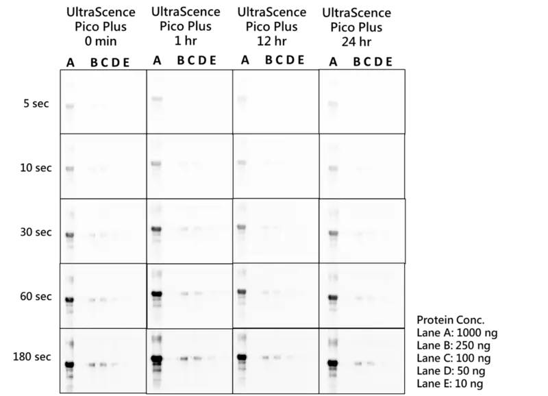
Figure 2. 24-hour stability test of UltraScence Pico Plus Western Substrate
The Luminol and Peroxide solution of UltraScence Pico Plus were mixed in a 1:1 ratio and having 400 uL treated on each blot after taken out from 4℃ for 1hr, 12 hrs and 24 hrs. The blots were simultaneously exposed for 5, 10, 30, 60, and 180 seconds using Chemlux SPX-600 Series digital imaging system. The EGFP (Enhanced Green Fluorescent Protein) was serially diluted for electrophoresis, after the proteins were transferred to the membrane, they were blocked and probed with GFP tag Rabbit PolyAb (Cat No. 50430-2-AP, 1:10,000), and followed by the Goat Anti-rabbit IgG/HRP secondary antibody (Cat No. SA00001-2, 1:10,000).
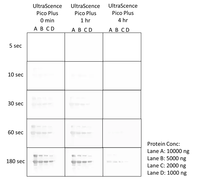
Figure 3. 4-hour signal duration with UltraScence Pico Plus Western Substrate
The EGFP (Enhanced Green Fluorescent Protein) was serially diluted for electrophoresis, after the proteins were transferred to the membrane, they were blocked and probed with GFP tag Rabbit PolyAb (Cat No. 50430-2-AP, 1:10,000), and followed by the Goat Anti-rabbit IgG/HRP secondary antibody (Cat no. SA00001-2, 1:10,000). Immerse the membrane in a mixed luminol and peroxide solution and check the signal at different times. The blots were simultaneously exposed for 5, 10, 30, 60, and 180 seconds using the Chemlux SPX-600 Series digital imaging system.
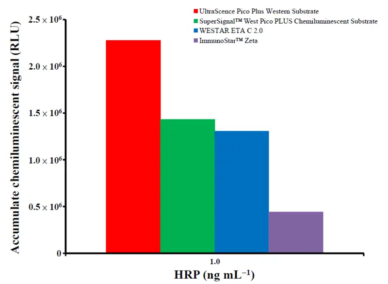
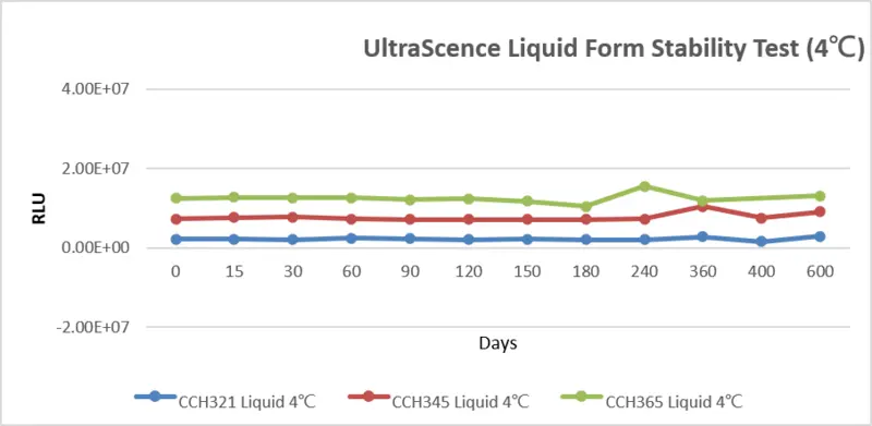
ELISA Application
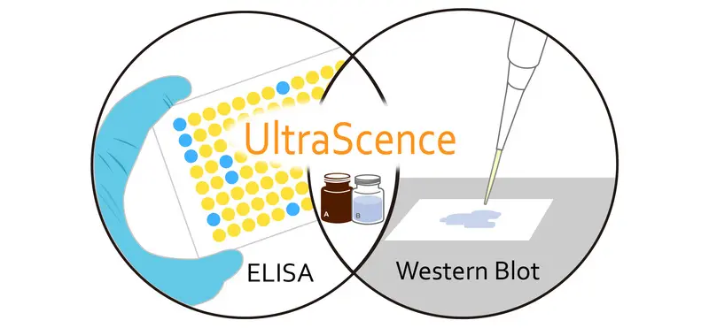
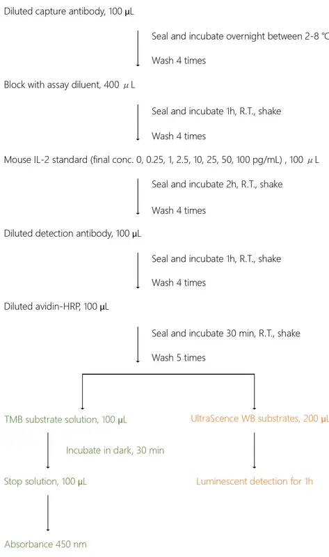
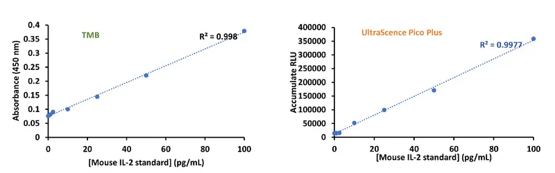
UltraScene WB substrates can also be applied as ELISA substrates
Frequently Asked Questions
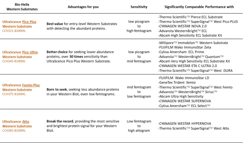
UltraScence ECL substrates series is compatible with the use from low picogram to low-femtogram level detections. Please kindly refer to the ECL selection guide of UltraScence Western substrate as the below table:
(a) BHCCH321B100ML, UltraScence Pico Plus Western Substrate 50ml+50ml
(b) BHCCH345B100ML, UltraScence Pico Ultra Western Substrate 50ml+50ml
(c) BHCCH375B100ML, UltraScence Femto Plus Western Substrate 50ml+50ml
(d) BHCCH385B100ML, UltraScence Atto Western Substrate 50ml+50ml