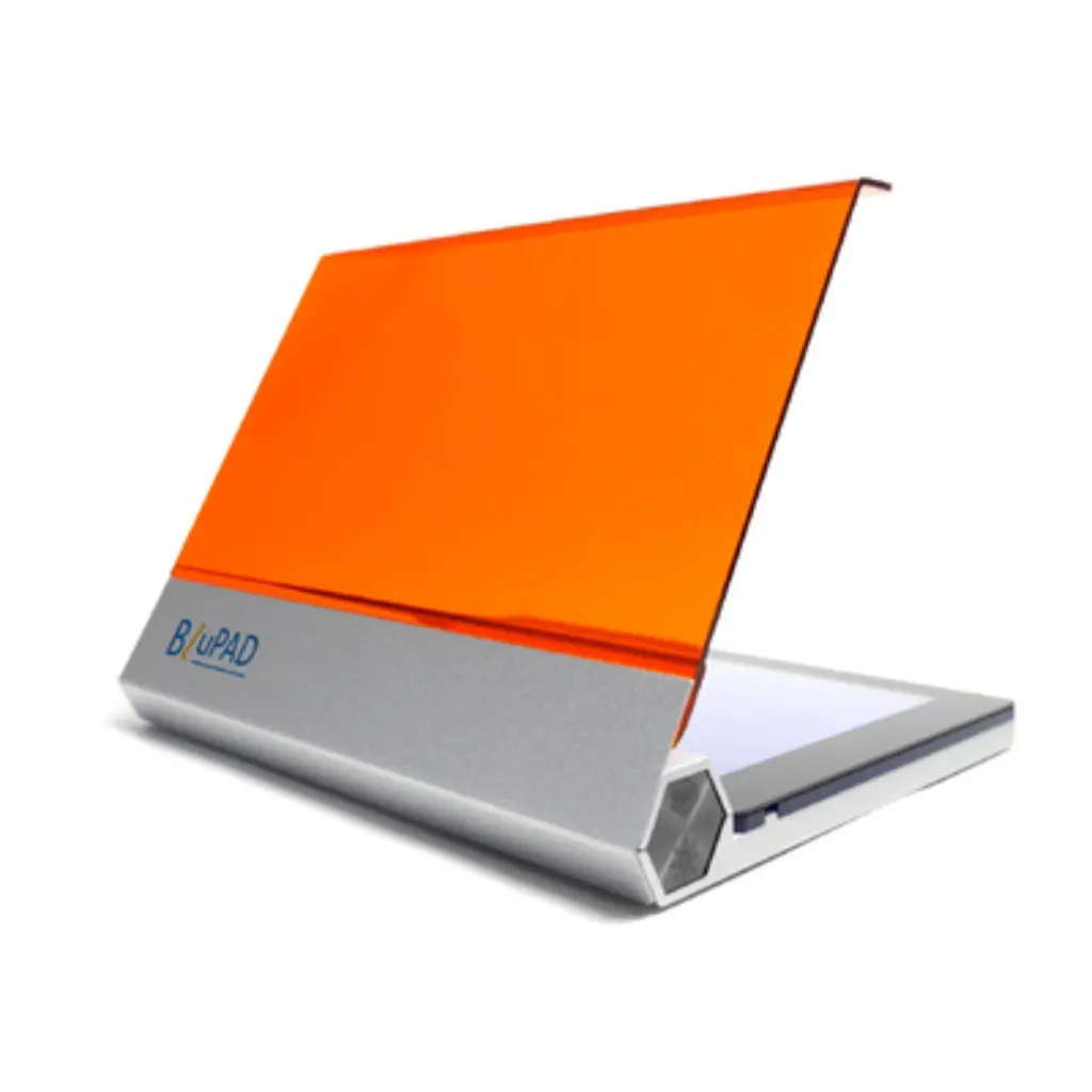
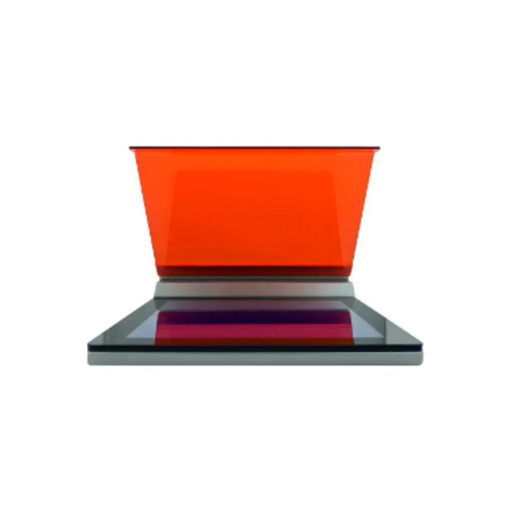
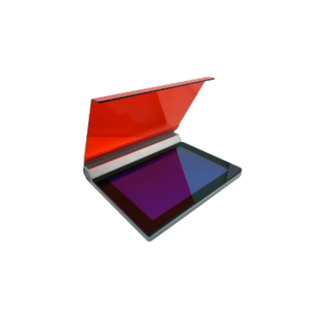
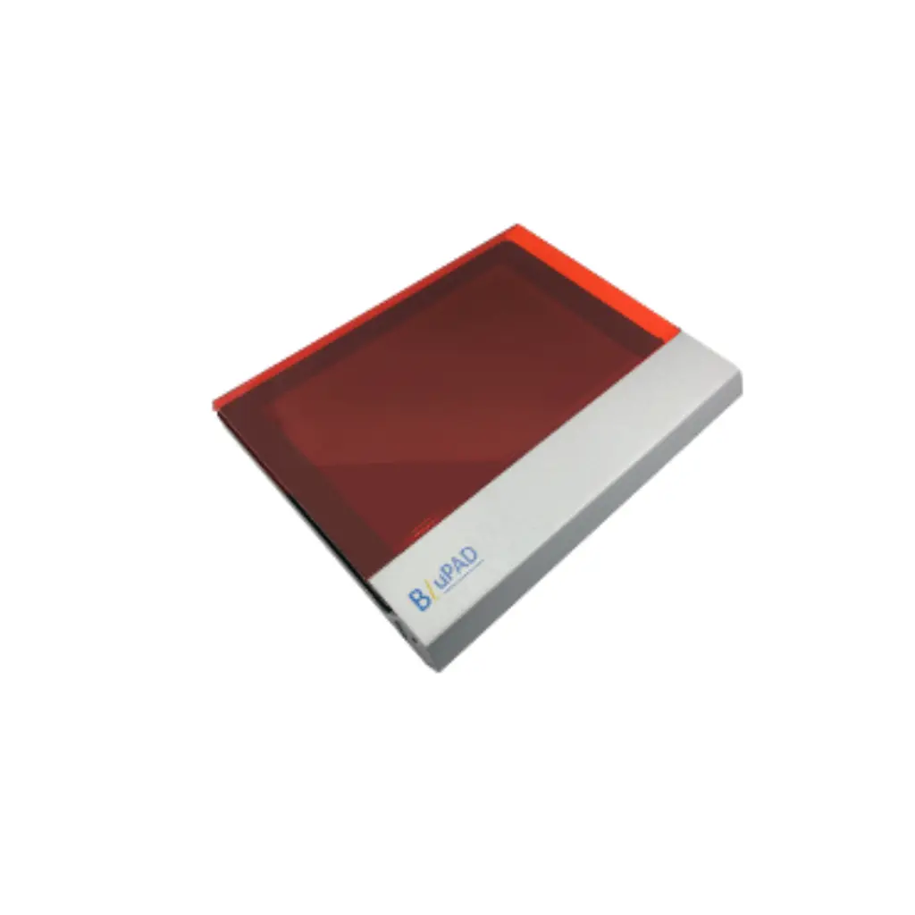
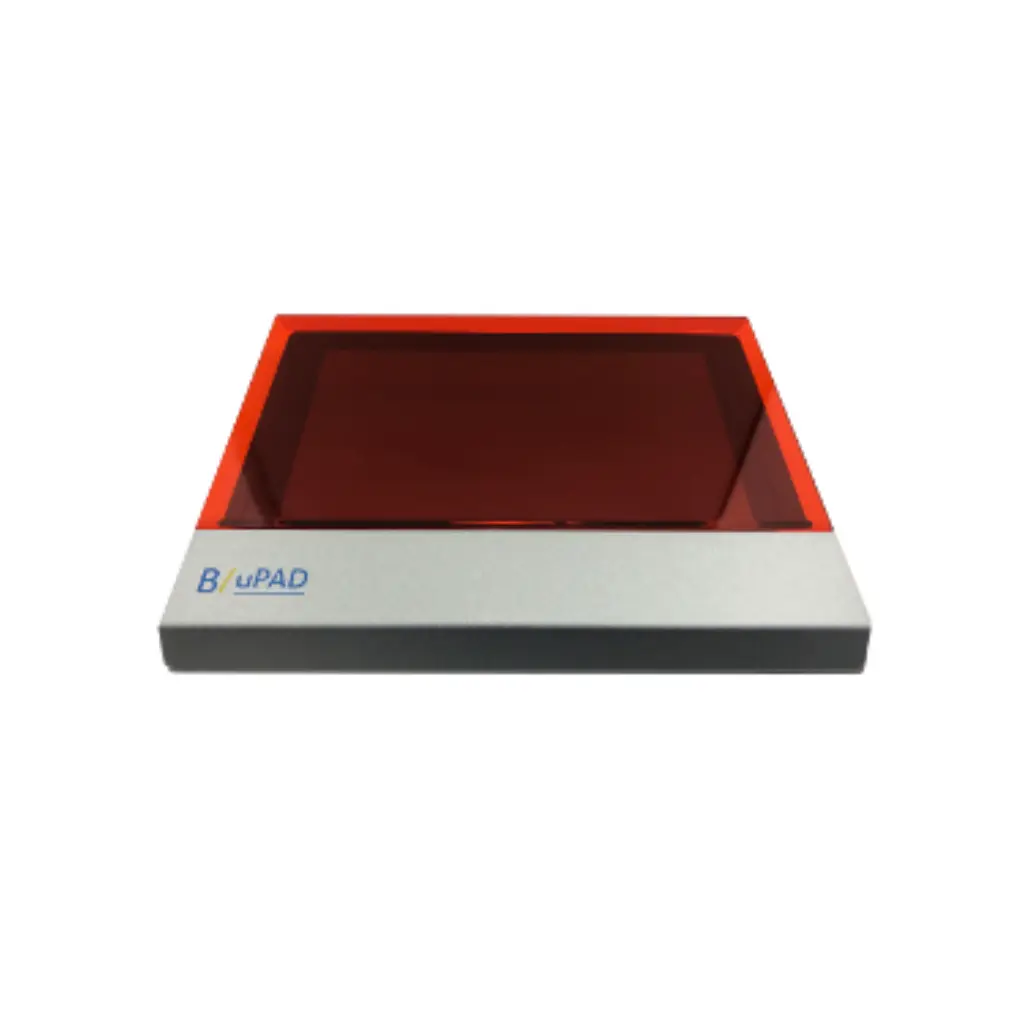
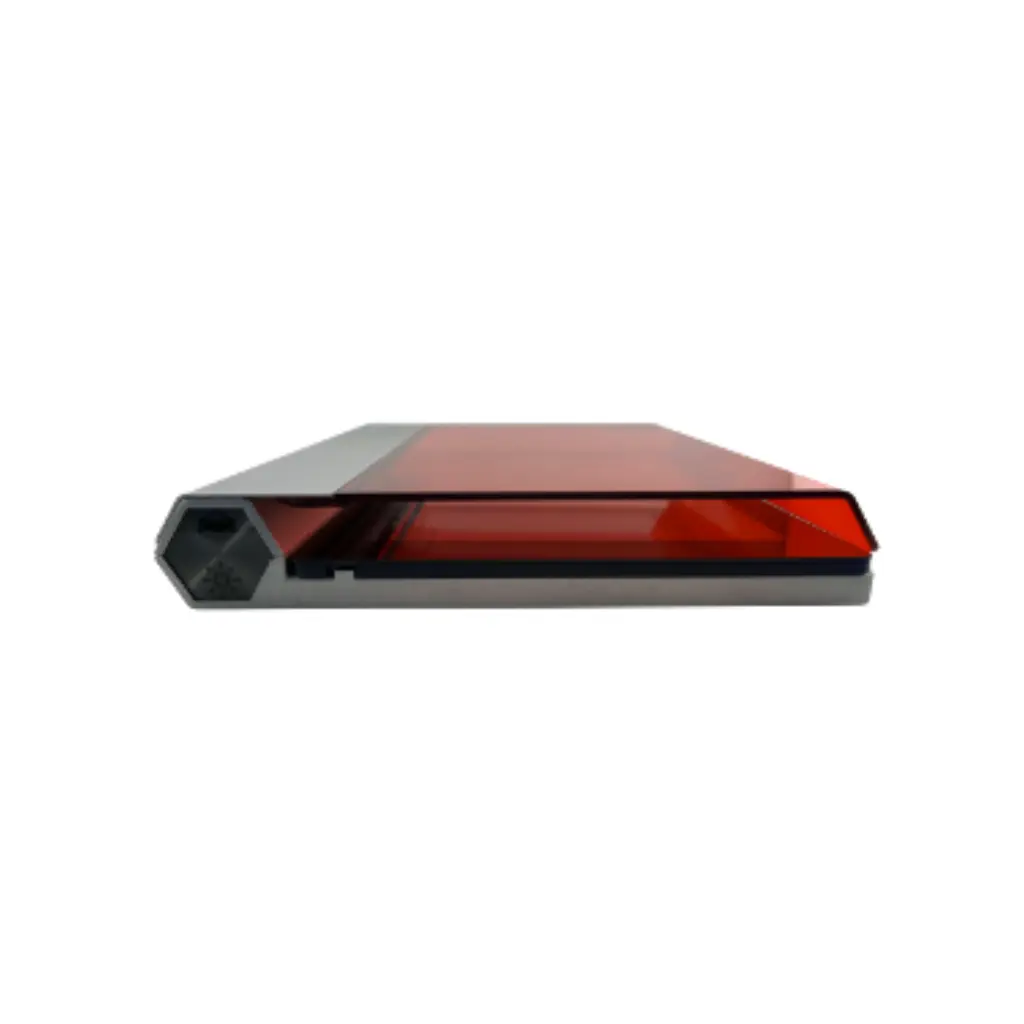
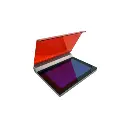
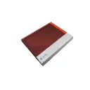
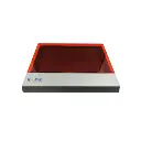
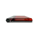
BluPAD Blue Light Transilluminator
A blue light transilluminator that shows you your DNA without destroying it
The light you use to view a gel impacts whether the DNA on it is still usable afterward. UV transilluminators damage the sample while you look at it, which shows up later as failed ligations and low cloning efficiency, and they burn unprotected eyes and skin in the process. The BluPAD excites fluorescent DNA stains with 470 nm blue LED light instead, so the bands glow, the DNA stays intact, and nobody needs a face shield.
A second, white light mode handles Coomassie and silver stained protein gels plus X-ray film viewing, making the BluPAD one compact LED transilluminator for both nucleic acid and protein work.
470 nm blue LED excitation with a 580 nm amber filter. No UV, no goggles.
White light mode for Coomassie and silver stained SDS-PAGE gels and X-ray film.
Compatible with SYBR Green, SYBR Gold, GelGreen, GelStar, SYPRO stains, and most fluorescent dyes on the market.
12 x 18 cm viewing area with three adjustable intensity levels.
LEDs rated beyond 30,000 hours, with automatic power-off after 5 minutes.
Runs from an external power bank for field work (power bank sold separately).

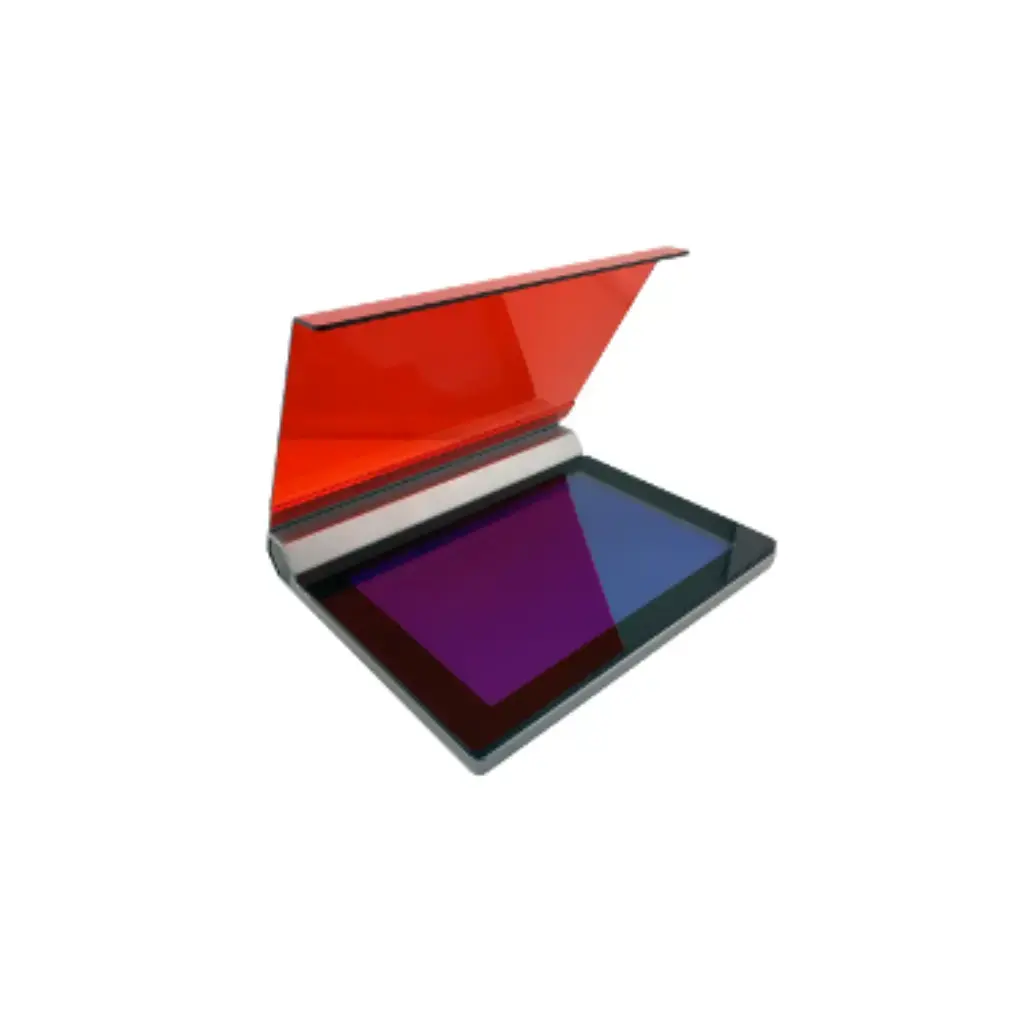
Why blue light beats UV for DNA gel viewing
UV light excites DNA stains well, and that is the only good thing it does at the bench. While you view or photograph the gel, the same UV exposure damages the DNA in it. If you plan to cut a band out for cloning, sequencing, or any downstream reaction, every second on a UV box costs you recovery. UV also requires protective gear, because the light that damages your sample damages your eyes and skin the same way.
Blue LED light at 470 nm excites modern fluorescent stains without either problem...
DNA stays intact during viewing and gel cutting, protecting downstream cloning and sequencing results.
No goggles or face shield required. The magnetic 580 nm amber filter screens the excitation light while you work.
LEDs switch on instantly, run cool, and hold their output past 30,000 hours. No warm-up, no bulb replacement schedule.
For labs still staining with ethidium bromide, blue light also opens the door to switching stains. Modern dyes like GelGreen and SYBR Safe were built as ethidium bromide alternatives, designed for blue light excitation, and safer to handle and dispose of.
One LED transilluminator for DNA gels, protein gels, and film
Blue light mode for stained DNA and RNA gels
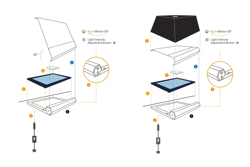
The 470 nm LED module excites fluorescent nucleic acid and protein stains for band checking, gel photography, and band excision. Three intensity levels let you match the light to the sample concentration: high for faint bands that would wash out, low for heavily loaded gels that need contrast.
Left : Gel Cutting / Right : Imaging
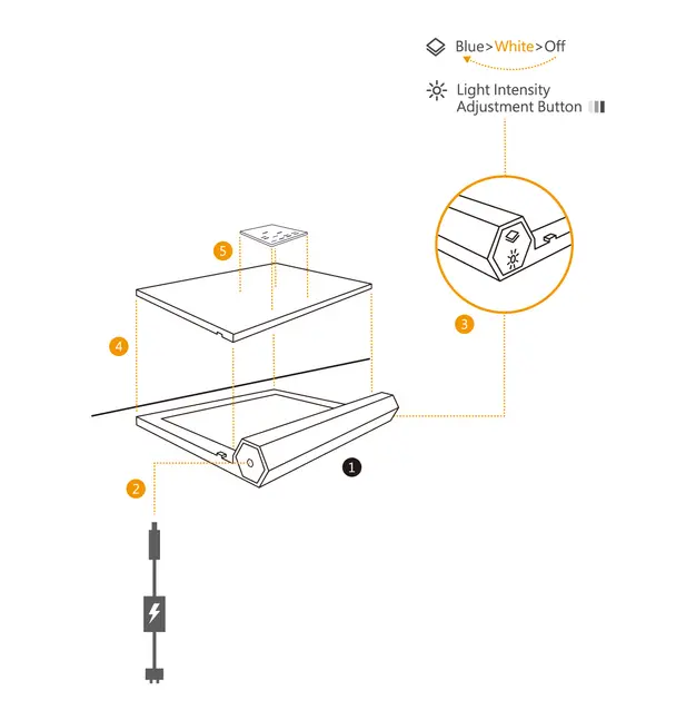
White light mode for Coomassie, silver stain, and X-ray film
The whole-wavelength white LED gives soft, uniform illumination from below for SDS-PAGE gels stained with Coomassie Blue or silver stain, where you need even light rather than excitation. The same mode works as a white light transilluminator for reading X-ray film at the bench.
Field mode for work away from the bench
The BluPAD connects to a standard external power bank, so gels can be checked at a field station or teaching site without a wall outlet. The power bank is sold separately.
Which stains work with the BluPAD
The blue light mode excites the fluorescent stains most labs already use...
 SYBR Green I and II, SYBR Gold, and SYBR Safe.
GelGreen and GelStar.
SYPRO Ruby, SYPRO Orange, and Coomassie Fluor Orange protein stains.
Bio-Helix stains including Novel Green, Novel Green Plus, Novel Juice, Primer Juice, and Nimble Juice.
Most other fluorescent DNA and protein stains on the market.
Ethidium bromide gels are visible too. EtBr has a secondary excitation peak between 400 and 570 nm, so bands appear under blue light at lower brightness than a UV box produces. Labs mid-transition can keep running EtBr gels while switching to a safer stain.
Do you need a gel documentation system or a transilluminator?
A gel documentation system is an enclosed cabinet with a built-in camera, filters, and analysis software. It earns its price when you need quantitative densitometry, chemiluminescent western blot imaging, or a standardized publication imaging workflow.
Most day-to-day gel work asks for less: run the gel, check the bands, photograph the result, cut out the fragment you need. A transilluminator covers all of that at a fraction of the cost of an imaging cabinet, and the BluPAD adds the white light mode a basic gel doc setup does not include. Many labs pair the two, using the imaging system for final documentation and a benchtop transilluminator for the everyday checking and cutting that would otherwise queue up behind it.
Blue light transilluminator specifications
Small details that matter on your bench
The amber filter attaches magnetically with no hinges, so there is nothing to snap off, and it lifts away in one motion for gel cutting.
Illumination comes from directly below the gel, so there are no reflections off the gel surface to wash out faint bands in photos.
Automatic power-off after 5 minutes protects the unit when someone walks away mid-experiment. It switches back on instantly.
The metal housing keeps the unit planted during gel cutting instead of sliding around the bench.
At 18.5 x 22 x 3 cm, it stores in a drawer and moves between benches, teaching labs, and field sites without a cart.
Common questions about blue light transilluminators
A transilluminator is a light box that shines light up through an electrophoresis gel from below. The stain bound to your DNA, RNA, or protein fluoresces or shows contrast under that light, making the bands visible so you can check results, photograph the gel, or cut bands out for downstream work.
UV light damages the DNA in the gel while you view it, reducing cloning and sequencing success from excised bands, and it requires eye and skin protection. Blue LED light at 470 nm excites modern fluorescent stains without damaging the sample or the person viewing it.
Yes. SYBR Safe was designed for blue light excitation, and it performs well on the BluPAD along with SYBR Green I and II, SYBR Gold, GelGreen, GelStar, and most other fluorescent stains on the market.
Yes. Ethidium bromide has a secondary excitation peak between 400 and 570 nm, so EtBr-stained bands are visible under the 470 nm blue light. They appear dimmer than under UV excitation at 300 nm, so increase the intensity setting and expect lower brightness on faint bands.
The viewing area measures 12 x 18 cm, which covers standard mini gels and most midi gel formats.
No. The 580 nm amber filter screens the blue excitation light during viewing, and because the source is blue LED rather than UV, gel cutting with the filter lifted does not expose you to harmful light.
Yes. Shoot through the amber filter with a phone or camera. The bottom-up illumination avoids surface reflections, and the three intensity levels let you set the contrast for the sample load you are imaging.
Yes. The white light mode illuminates SDS-PAGE gels stained with Coomassie Blue or silver stain, and the blue light mode excites fluorescent protein stains like SYPRO Ruby, SYPRO Orange, and Coomassie Fluor Orange.
Confirm the blue light source is selected rather than white light, check that the protective films on the white plate and blue tempered glass were removed before first use, and raise the intensity setting. Very low sample concentrations may need a higher gel load to produce visible bands.
Yes. The BluPAD connects to an external power bank for field stations and teaching sites. The power bank is sold separately.